By Dr Thomas Gorochowski and insights from PhD student Janine McCaughey and Research Associate Ulrike Obst.
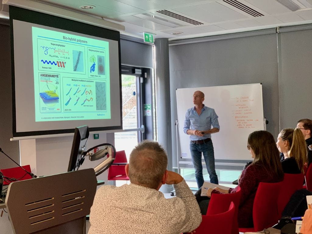
On the 2nd October 2019, Professor Nico Sommerdijk visited Bristol for the monthly BBI seminar and provided a picture of the hidden nanoscale world underpinning biology. Nico, who describes himself as a synthetic organic chemist who got carried away when introduced to the expanding capabilities of electron microscopy (EM), has since then never looked back. His work spans the development and application of new EM imaging and microscopy methods to not only image molecular assemblies in a single static pose, but also to record movies that help unravel the steps involved in their self-assembly.
The seminar focused on the mineralisation of collagen fibril that act as the basic building block of our bones. The nanoscopic dimensions and complex, hybrid composition of mineralised collagen makes it difficult to examine. Trickier still is monitoring the multi-step process that collagen goes through to mineralise, as it forms not only at a billionth of a meter in scale, but also in an intricate, aqueous environment.
Why is understanding this process important?
Understanding the process of collagen mineralization could enable the development of new treatments for bone defects and disorders of bone mineralization, such as rickets and hypophosphatasia.
Nico highlighted that “it will also offer new opportunities for the design of new bio-inspired materials”. This can be in the form of an in vitro unit that can be biologically engineered to form mineralized collagen to precise specifications. Unravelling the minute process of mineralization could therefore benefit many branches of science, from medicine and pharmaceuticals to bioengineering.
What did the audience think?
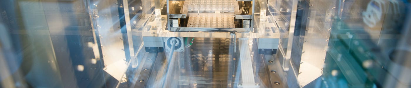
Janine McCaughey from the School of Biochemistry explained that “Nico presented an interesting approach to enabling live cell TEM in form of a liquid chamber. This allows for imaging of dynamic processes in high resolution compared to cryoTEM that requires one sample per timepoint and therefore only delivers very limited insight into such processes”.
Ulrike Obst, a Research Associate from Cellular and Molecular Medicine, was amazed by the sheer scope of the work. “It was fascinating to find out about so many different electron microscopy techniques, and especially how they can be combined to observe dynamic biological processes at a nanoscale!”
Ulrike was also amused by this idea of “having a mini-aquarium in a microscope”, where a tiny pocket of water allows for molecular self-assembly to be watched in real-time. “It is crazy that such things are possible. It’s like a window into another world.”
Interested to find out more?
As part of his recent ERC Advanced Grant award titled “A Google Earth Approach to Understanding Collagen Mineralization”, Nico recently moved to the Radboud Institute for Molecular Life Sciences in Nijmegen, Netherlands (https://www.radboudumc.nl/en). As part of this project, his group will try to figure out how collagen is assembled by developing and combining methods able to image from the nano- to cellular-scales, providing an unprecedented understanding of this crucial biological process.