There will be much more to come throughout 2023, and we are looking forward to beginning the year with the BrisEngBio Annual meeting, and BrisEngBio Connect Partnership and Networking Event.
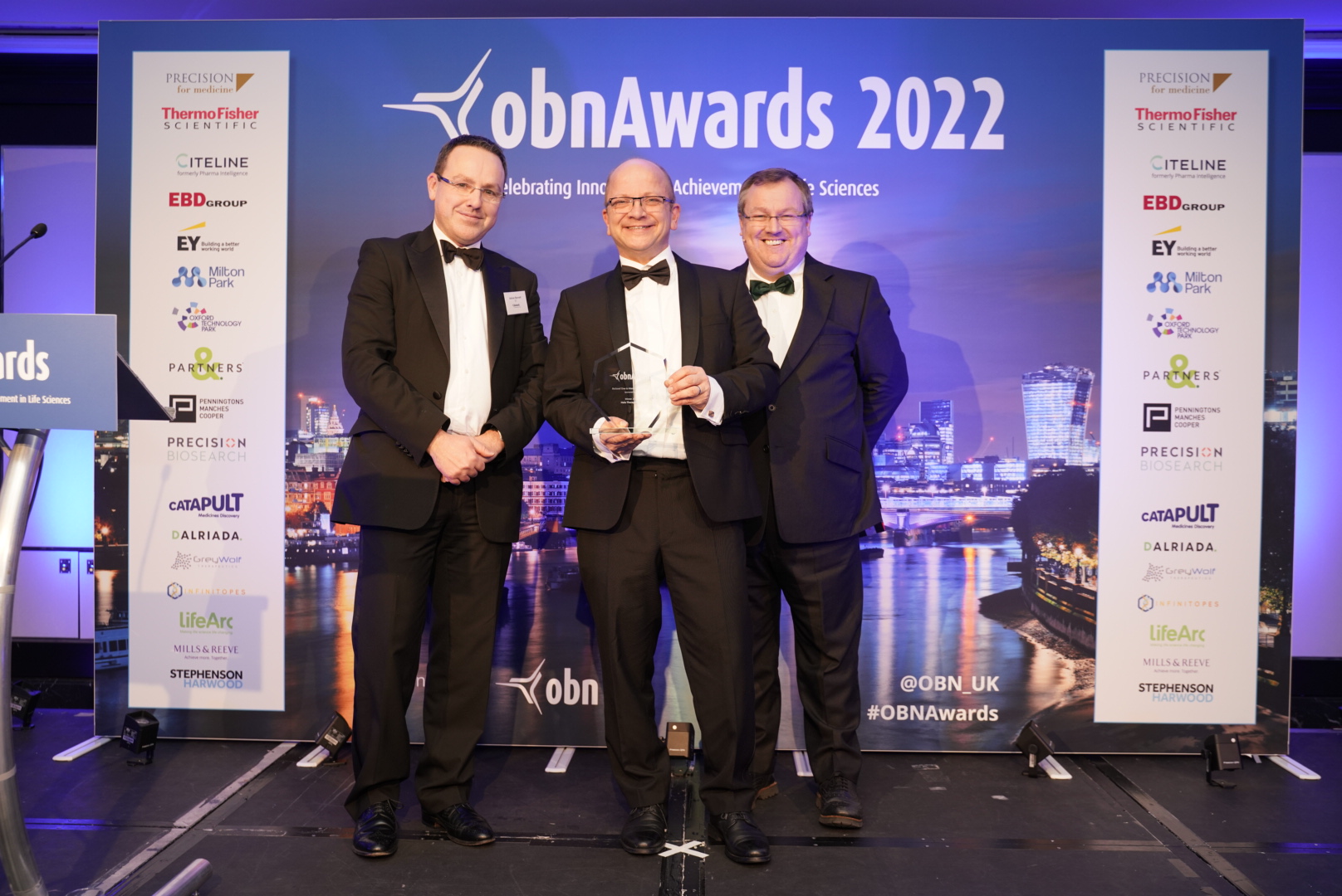
Congratulations to the team at Halo Therapeutics, who have won the ‘BioSeed ‘One to Watch’ Award for Therapeutics’ at the OBN Awards. Halo is developing pan coronavirus antivirals for home use by COVID-19 patients, based on groundbreaking discoveries made during the lockdowns. The award was collected by Prof Imre Berger, who is co-founder and CSO of Halo, and a Co-Director of the Bristol BioDesign Institute. Halo was founded on IP generated through BrisSynBio.
Prof Imre Berger collecting the OBN Award on behalf of Halo Therapeutics (Photo: OBN UK)
This technology relies on the ability of the CRISPR-Cas13a protein to detect specific sequences of RNA when coupled with a crRNA guide. crRNA guides can be designed to detect any unique target sequence. When the protein-RNA complex finds the target sequence, it becomes activated and begins to cleave any RNA molecules in its path, including RNA ‘reporter’ probes which ‘light up’ when cleaved.
The SHERLOCK system uses this approach to indicate the presence or absence of specific RNA sequences in samples. Please see the original publications for a schematic explaining this system in more detail [Kellner et al., (2019); Gootenberg et al., (2017)].
Producing Cas13a, a protein that can detect specific sequences of RNA
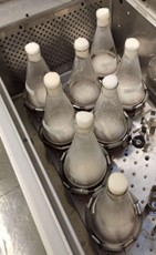
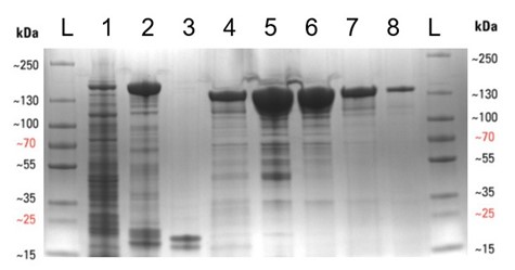
We began by transforming E. coli with the required plasmid (Addgene #90097) and then grew 8 litres of bacterial culture expressing the protein overnight (see below), before lysing the cells to release the protein. The Cas13a was then purified by affinity, ion-exchange, and finally size-exclusion chromatography methods, to yield a purified protein. This is especially important for the SHERLOCK assay, as any RNase contamination can give false positive results. The protein was then concentrated to 2 mg/mL, aliquoted and frozen at -80°C. The protein purification gel below displays the various stages of the expression and purification process. Thanks to the D40 protein gurus (Arthur, Alan and Zac, to name but a few) for all their help and advice during the purification.
Expression and purification of the Cas13a protein. (Left) The shake flasks used to grow the E. coli to express the Cas13a. (Right) SDS-PAGE protein purification gel displaying the various stages of the protein purification procedure, with protein purity increasing from left to right. (A more detailed figure is displayed at the end of this blog).
Designing crRNA target sequences
With the biosensor protein in hand, the team designed some genetic sequences that guide the protein to the target RNA, if it is present. Two computational biology tools were used: ADAPT [Metsky et al., (2022)] and BLASTN. Firstly, ADAPT was used to identify a target sequence unique to a particular organism. A house-keeping gene (rpoB) was used as a starting point, since it is commonly used as a molecular marker in microbial ecology studies [Case et al., (2017)]. Since our preliminary studies concerned the detection of a particular microorganism within mock microbial community DNA, we ran ADAPT against the rpoB sequences of other species in the community. The absence of the target sequence in other bacterial species was confirmed using BLASTN. The designed crRNA were ordered as DNA templates and then expressed using in vitro transcription, as per the SHERLOCK protocol [Kellner et al., (2019)].
Testing the biosensors
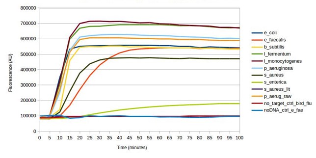
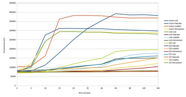
Mock microbial community DNA was used to validate the biosensor. crRNA sequences were designed and synthesised for all microorganisms found in the mock microbial community. Every design successfully gave a positive result (see below), detecting the target in the mock community sample, as intended. Furthermore, testing without the DNA present, and testing a target (bird flu) not present in the community, both gave a negative result. Some of the SHERLOCK reagents are particularly expensive and so after this first test, the assay was optimised to reduce reagent input, whilst maintaining adequate fluorescence from a positive result (results not shown). This reduced the fluorescent output of a positive result from the mock community from around 600,000 AU to around 300,000 AU.
Testing SHERLOCK biosensors against mock community DNA
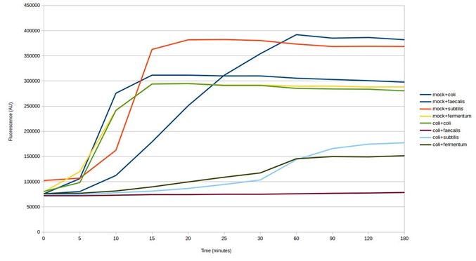
The next step was to test the biosensors for cross-reaction using E. coli DNA. The results show some of the biosensors ‘light up’ in response to E. coli DNA, giving a false positive result (see below). This is despite being designed to avoid targeting other microbes in the mock microbial community. Whilst the strength of the response is weaker, testing further biosensor design iterations would be necessary to identify those that are specific to particular microorganisms.
Testing SHERLOCK biosensors against mock community and E. coli DNA
Finally, the team tested four of the biosensors against two DNA samples that had been extracted from soil. Three of the four biosensors gave a positive result for both samples (see below). The measured signal was weaker than detection of species in the mock community. This could be a result of the lower concentration of targets in the environmental DNA samples. Environmental microbiomes contain many thousands of microorganisms, which would make detection of specific species challenging. These results could be confirmed by DNA sequencing the environmental DNA samples.
Testing SHERLOCK biosensors against mock community environmental DNA (D2 & S2)
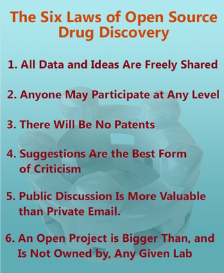
The six laws of open source drug discovery are equally relevant to open source synthetic biology research (image reproduced from Todd et al., with permission)
All considered, these initial results show that further optimisation is required to achieve the main goal of this ambitious project: to allow the detection of specific bacterial species in water-based samples. Whilst the biosensors detected the target species, they appeared to be susceptible to activation by RNA from other species, resulting in false positive results. This living planet is estimated to be home to one trillion microbial species, making it tricky, but not impossible, to design biosensors for the detection of particular microorganisms.
We are sharing these results to encourage a mindset of open source research, for which six guiding principles have been proposed [Todd, (2016)].
We would like to thank the SynBio CDT for the funding that facilitated this project, the D40 and Biocompute labs for the use of lab space and equipment, and Kathleen Sedgley for the help in setting up the project.
***
Supplementary
Expression and purification of the Cas13a protein. SDS-PAGE protein purification gel displaying the various stages of the protein purification procedure, with protein purity increasing from left to right. Lanes are as follows: L – protein ladder, 1 – Cleared cell lysate post sonication, 2 – Streptactin resin post binding, 3 – Streptactin resin post cleavage, 4 – Flow-through post cleavage, 5 – Concentrated sample post IEC , 6 – Concentrated sample post SEC, 7 – Diluted Cas13a aliquot (5 µL), 8 – Diluted Cas13a aliquot (1 µL). The molecular weight of SUMO-Cas13a is 155 kDa and Cas13a (post SUMO cleavage) is 139 kDa.
***
References:
Case, R. J. et al. (2007) “Use of 16S rRNA and rpoB Genes as Molecular Markers for Microbial Ecology Studies”, Applied and Environmental Microbiology, 73(1), pp. 278–288. Available at: doi.org/10.1128/AEM.01177-06.
Gootenberg, J. S. et al. (2017) “Nucleic acid detection with CRISPR-CAS13A/C2C2”, Science, 356(6336), pp. 438–442. Available at: doi.org/10.1126/science.aam9321.
Kellner, M. J. et al. (2019) “Sherlock: Nucleic acid detection with CRISPR nucleases”, Nature Protocols, 14(10), pp. 2986–3012. Available at: doi.org/10.1038/s41596-019-0210-2.
Metsky, H. C., et al. (2022). “Designing sensitive viral diagnostics with machine learning”, Nature Biotechnology, pp. 1–9. Available at: doi.org/10.1038/s41587-022-01213-5.
Todd, M. H. (2019). “Six Laws of open source drug discovery”, ChemMedChem, 14(21), pp. 1804–1809. Available at: doi.org/10.1002/cmdc.201900565.
Ben Hardy, a Research Associate in the School of Biochemistry at the University of Bristol, won a prize for his poster at the Synthetic Biology UK conference (7-8 November 2022).
Summarising work from his PhD, Ben’s poster (Computational Design of a de novo Transmembrane Cytochrome) highlights the successful design of an artificial protein capable of electron transport within membranes – such a protein is an essential component for constructing novel bioenergetic complexes within cells.
This was Ben’s second poster success this year – he won first prize at the Advances in Protein Folding, Evolution & Design conference (April 2022) for the poster: ‘Computational design of Bioenergetic Membrane Proteins’ (pictured).
Red blood cells that have been grown in a laboratory have now been transfused into another person in a world first clinical trial led by a UK team including University of Bristol researchers.
This is the first time in the world that red blood cells that have been grown in a laboratory have been given to another person as part of a trial into blood transfusion.
If proved safe and effective, manufactured blood cells could in time revolutionise treatments for people with blood disorders such as sickle cell and rare blood types. It can be difficult to find enough well-matched donated blood for some people with these disorders.
The trial is studying the lifespan of the lab grown cells compared with infusions of standard red blood cells from the same donor. The lab-grown blood cells are all fresh, so the trial team expect them to perform better than a similar transfusion of standard donated red cells, which contains cells of varying ages.
Additionally, if manufactured cells last longer in the body, patients who regularly need blood may not need transfusions as often. That would reduce iron overload from frequent blood transfusions, which can lead to serious complications.
The trial is the first step towards making lab grown red blood cells available as a future clinical product. For the foreseeable future, manufactured cells could only be used for a very small number of patients with very complex transfusions needs. NHSBT continues to rely on the generosity of donors.
Two people have so far been transfused with the lab-grown red cells. They were closely monitored and no untoward side effects were reported. They are well and healthy. The identities of participants infused so far are not currently being released, to help keep the trial ‘blinded’.
The amount of lab grown cells being infused varies but is around 5-10mls – about one to two teaspoons.
Donors were recruited from NHSBT’s blood donor base. They donated blood to the trial and stem cells were separated out from their blood. These stem cells were then grown to produce red blood cells in a laboratory at NHS Blood and Transplant’s Advanced Therapies Unit in Bristol. The recipients of the blood were recruited from healthy members of the National Institute for Health and Care Research (NIHR) BioResource.
A minimum of ten participants will receive two mini transfusions at least four months apart, one of standard donated red cells and one of lab grown red cells, to find out if the young red blood cells made in the laboratory last longer than cells made in the body.
Further trials are needed before clinical use, but this research marks a significant step in using lab grown red blood cells to improve treatment for patients with rare blood types or people with complex transfusion needs.
Co-Chief Investigator Ashley Toye, Professor of Cell Biology at the University of Bristol and Director of the NIHR Blood and Transplant Unit in red cell products, said: “This challenging and exciting trial is a huge stepping stone for manufacturing blood from stem cells. This is the first-time lab grown blood from an allogeneic donor has been transfused and we are excited to see how well the cells perform at the end of the clinical trial.”
Co-Chief Investigator Cedric Ghevaert, Professor in Transfusion Medicine and Consultant Haematologist the University of Cambridge and NHS Blood and Transplant, said: “We hope our lab grown red blood cells will last longer than those that come from blood donors. If our trial, the first such in the world, is successful, it will mean that patients who currently require regular long-term blood transfusions will need fewer transfusions in future, helping transform their care.”
Dr Rebecca Cardigan, Head of Component Development NHS Blood and Transplant and Affiliated Lecturer at the University of Cambridge said: “It’s really fantastic that we are now able to grow enough red cells to medical grade to allow this trial to commence, we are really looking forward to seeing the results and whether they perform better than standard red cells.”
John James OBE, Chief Executive of the Sickle Cell Society, said: “This research offers real hope for those difficult to transfuse sickle cell patients who have developed antibodies against most donor blood types. However, we should remember that the NHS still needs 250 blood donations every day to treat people with sickle cell and the figure is rising. The need for normal blood donations to provide the vast majority of blood transfusions will remain. We strongly encourage people with African and Caribbean heritage to keep registering as blood donors and start giving blood regularly.”
Dr Farrukh Shah, Medical Director of Transfusion for NHS Blood and Transplant, said: “Patients who need regular or intermittent blood transfusions may result develop antibodies against minor blood groups which makes it harder to find donor blood which can be transfused without the risk of a potentially life-threatening reaction. This world leading research lays the groundwork for the manufacture of red blood cells that can safely be used to transfuse people with disorders like sickle cell. The need for normal blood donations to provide the vast majority of blood will remain. But the potential for this work to benefit hard to transfuse patients is very significant.”
A study led by the labs of Prof Steve Mann (Chemistry) and Prof Paul Martin (Biochemistry) at the University of Bristol has demonstrated a new potential anti-cancer therapy that boosts the body’s inflammatory response using miniature artificial particles called protocells. This discovery is documented in an article published in Advanced Science, which specialises in interdisciplinary studies.
Our inflammatory cells, macrophages and neutrophils in particular, have remarkable cancer surveillance capacities, and therefore they act as “vigilants” to detect any cancer cell arising in the body. However, when these immune cells encounter the malignant cells, rather than killing them, they generally feed them with nutrients, which favours cancer growth. In this study, we developed a novel therapeutic system to reprogram immune cells away from cancer nurturing and towards cancer killing.
To achieve this, we injected miniature artificial protocells into the bloodstream of zebrafish, which are translucent and thus allow us to live image protocell dynamics and cell-cell interactions in real time. We observed that protocells were selectively taken up by inflammatory cells which were then reprogrammed, by specific cargoes loaded in the protocells, to make them more effective at killing cancer. The “reprogramming” cargo packaged in protocells was a miR223 inhibitor, anti-miR223, which maintains a pro-inflammatory/anti-cancer state in inflammatory cells, otherwise repressed by the presence of endogenous miR223. We showed that protocell-mediated reprogramming of the immune response led to reduced cancer cell proliferation and melanoma shrinkage in zebrafish.
In collaboration with Prof Ash Toye’s lab (Biochemistry), we established an in vitro assay with human macrophages supplemented with protocells. Our results showed that the protocells were able to promote anti-cancer reprogramming in human macrophages, too, which suggests that this novel protocell system may be a promising cancer immunotherapy strategy against human melanomas, and possibly also for other cancers with a pro-inflammatory vulnerability.
The research is explained in this scribble video:
Having demonstrated the ability of protocells to deliver cargoes to tumour-associated leukocytes and enhance their anti-cancer capacities, the Mann and Martin groups now plan to expand the therapeutic application of the protocell system beyond cancer and test the feasibility of using protocells to modulate the inflammatory response to unresolved/chronic wounds in order to improve wound healing in these clinically relevant inflammatory conditions. This research is currently ongoing and is supported by funding from BrisEngBio.
Macrophage Reprogramming with Anti-miR223-Loaded Artificial Protocells Enhances In Vivo Cancer Therapeutic Potential
Paco López-Cuevas, Can Xu, Charlotte E. Severn, Tiah C. L. Oates, Stephen J. Cross, Ashley M. Toye, Stephen Mann, Paul Martin Adv. Sci. 2022, 2202717, https://doi.org/10.1002/advs.202202717
Zentraxa, founded on IP generated through BrisSynBio, specialises in the design, production and testing of novel biomaterials. It has received £320k funding for commercial concept development of both medical adhesives and personal care ingredients that could have use in medicine, for example, skin bonding and wound care, or in personal use, such as skin and hair products.
Glaia, which benefitted from BrisSynBio commercialisation funding and dedicated innovation support, has secured £1 million in new investment to develop its carbon-based technology, the ‘sugar dots’, which increases crop yields and reduces emissions from crops by 30% when applied to the plants.
The Universities of Bristol and Edinburgh are collaborating on the Enzymatic Photocatalysis project, which will be led by Prof Nigel Scrutton at University of Manchester. This project will “apply a cyclical design-build-evaluate-learn approach to discovering the generalisable principles of photo-biocatalysis.”
The Bristol team will design completely synthetic proteins that trap light and funnel this energy into new enzyme-like activities for generating molecules that are otherwise difficult to make synthetically or biochemically.
Overview
Two BBSRC-funded post-doctoral research associate positions are available immediately to work in Dek Woolfson’s Peptide Design and Assembly group in the Schools of Chemistry and Biochemistry at the University of Bristol. Experience in one or more of peptide chemistry, protein biochemistry, de novo peptide/protein design, and the structural characterization of peptides and proteins would be an advantage. However, such experience is not essential, as we are looking for enthusiastic and talented researchers in the chemical/biochemical sciences who are interested in pursuing careers in peptide/protein design and its application in chemical and synthetic biology.
Post 1: De novo protein design in cells
This three-year post is joint with Dr Mark Dodding’s group (Biochemistry, Bristol). It builds on recent work between the Woolfson and Dodding groups (Cross et al.Cell Chem Biol(2021) DOI: 10.1016/j.chembiol.2021.03.010; Rhys et al.Nature Chem Biol(2022) DOI: 10.1038/s41589-022-01076-6). The project aims to design motor proteins from the bottom up to operate in eukaryotic cells.
Environment The Woolfson lab has purposed-built office space for computational work and laboratories for peptide chemistry, protein biochemistry, biophysics, protein crystallization, and cell biology. In addition, through Chemistry, Biochemistry, and the Bristol BioDesign Institute, the group has walk-up access to mass spectrometry, light and electron microscopy, and other facilities. The current group comprises 16 people with a balance of PhD and post-doctoral researchers from diverse backgrounds from around the world, which fosters a supportive, inclusive, and cutting-edge approach to peptide-design research.
 or of the Max Planck Bristol Centre for Minimal Biology, has talked to Mike Stiles about